Nowadays, an increasing number of veterinary diagnostic tools are available on the market, providing clear images, accurate measurements and quantified data. The first veterinary support tools were of course imaging techniques, with the arrival of radiography more than a century ago, but recent innovations have also challenged traditional practices.
In this article, we will review the different tools available for veterinarians to support their expertise and diagnosis.
Proximity imaging techniques
Since the end of the 1970s, imaging techniques have greatly evolved. They provide key information to veterinarians in the investigation of locomotor disorders.
In addition to allowing for a more informed diagnosis, they have enabled better documentation of lesions and ensured the best information/cost ratio and tolerance for the horse.
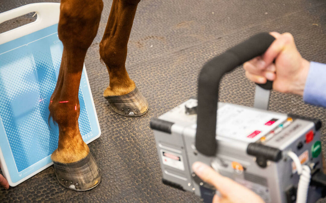
Digital radiography
During a veterinary examination of a horse, an X-ray of the bones and joints has almost become a routine procedure. It is even fair to say that it is now a requirement during purchase visits. Its application enables, among other things, the detection of physical anomalies that are not visible to the naked eye, to verify the absence of degenerative lesions of the joints or the detection and prediction of bone lesions.
This technique, which appeared more than a century ago, still has its limitations: poor differentiation of soft tissues (for which it must be combined with ultrasound) and poor sensitivity to certain bone lesions such as contusions and fatigue fractures at an early stage (which require the use of scintigraphy or MRI) (Denoix and al, 2004).
")
Ultrasound
Ultrasound has revolutionized the documentation of the causes of lameness in horses since the 1990s. It has naturally become the technique of choice for the diagnosis of tendonitis because it allows for the examination of soft tissues, particularly tendons and ligaments. It has brought to light a large number of previously unknown conditions, while also putting radiographic concepts into perspective, particularly in foot lameness.
By combining probes, ultrasound offers the additional advantage of evaluating all areas of the horse’s body (including endocavity: transrectal ultrasound) with the exception of spaces covered by bone (e.g., cranial cavity) or air (e.g., mediastinum occluded by the lungs). Like radiography, ultrasound is generally very well tolerated and non-invasive, and can be performed with small, easily transportable machines. The development of ultrasound-guided injections has also revolutionized the treatment of axial regions and proximal limb joints. (Denoix, 2000).
Thermography
Using infrared radiation, thermography provides a cartography of body areas. Because of its sensitivity to numerous artifacts (external temperature, skin and hair coat condition, etc.), it is more a technique for documenting the evolutionary and inflammatory stage of lesions than a diagnostic technique. It is useful in monitoring the healing of tendon injuries but requires perfectly standardized conditions of implementation.

Medical imaging techniques
Scintigraphy
Since the 1970s, bone scintigraphy has allowed the localisation of bone lesions in lameness of unknown origin. This technique begins with the intravenous injection of a very slightly radioactive product: Technecium 99m, linked to a bisphosphonate. Once injected, the product will fix itself on the whole skeleton of the horse and especially on the skeletal sites under construction or reconstruction (areas of bone inflammation or increased mechanical stress). Then, a gamma-camera placed in contact with the various anatomical regions of the horse, will detect the radioactivity emitted by the horse.
In addition to the ossification areas on the fertile side of the epiphyseal cartilages in young horses, the fixation points allow the detection of bone lesions induced by biomechanical constraints (contusions, fatigue fractures), bone remodeling associated with degenerative arthropathies or more rarely septic foci and, in exceptional cases, tumors.. To summarize, scintigraphy allows for the creation of a panoramic view of the horse’s potentially painful skeletal regions. (Denoix et Audigié, 2004).
Horses are isolated in stalls for more than 48 hours after the exam because it takes an average of 60 hours for the radioactive product to be eliminated by the animal. It should be noted that this tool is not particularly sensitive to soft tissue lesions and cannot detect tendon lesions.
Magnetic Resonance Imaging – MRI
The first MRI images on a living horse were performed in 1997 at the University of Washington. It was a few years later, in 2002, that MRI on a standing horse was developed. This imaging technique is now a reference thanks to its numerous advantages: a very good anatomical representation of various formations, bones and soft tissues, high sensitivity to various pathological processes (inflammation, bone sclerosis, tissue necrosis) (Tapprest et al., 2003)… And, the ability to perform slices in the three planes of space. It is therefore particularly useful for the early detection of fatigue fractures in racehorses or to diagnose bone inflammations not visible in close-up imaging in sport horses. MRI can be performed on a horse under general anesthesia, or on a standing horse.
Scanner
The scanner is a high-resolution sectional imaging technique based on X-rays. The first images were taken in the 1980s, still at the University of Washington. This tool allows, among other things, to obtain more precise images than with an X-ray thanks to millimeter cuts and offers the possibility of 3D reconstruction of bones and joints. During an examination, the scanner can be performed in different ways: under general anesthesia of large body regions or on a sedated horse for the head, especially the teeth and sinuses.
Connected tools
In recent years, connected devices have gained importance in the sports sector, both for performance monitoring and health management, and are gradually being integrated into the equine industry.
Connected sensors
Connected sensors allow considering new practices: veterinary telemedicine. These sensors, synchronized to an application or a platform, allow to follow and analyze the horse’s training without any geographical or temporal limit. By gathering numerous data such as heart rate, cadence, amplitude, fitness level or recovery capacity… These make it possible to diagnose the horse in real working conditions.
Exercising ECG
The embedded electrocardiogram is a real strength in veterinary diagnosis. Like the connected sensors, it allows to collect the horse’s ECG, directly during the effort, even at high speed. In a logic of investigation of underperformance, or longitudinal monitoring, these tools could revolutionize equine medicine, and thus preserve the physical integrity of athletic horses. A sensor like EQUIMETRE VET allows to visualise the horse’s ECG live via a mobile application, allowing for the detection of arrhythmias while working on a track or on a treadmill.
Dynamic Respiratory Scope
The Dynamic Respiratory Scope (DRS) is a tool of choice for the detection and management of some upper respiratory tract diseases. It is a portable endoscope, which attaches directly to the horse to provide a view of the airway during work. The advantage: detect a dynamic abnormality affecting the horse’s throat – an abnormality that appears during effort – and therefore not visible when the horse is at rest.
Locomotion quantification tools
Locomotion quantification systems allow to obtain precise and measurable data about locomotor asymmetries of the horse. These devices support veterinarians during a dynamic clinical examination and allow them to collect data invisible to the naked eye, with millimeter precision. A tool such as EQUISYM can be used in many ways, both in the diagnosis of complex lameness and in making comparisons before and after anesthesia or treatment.
CONCLUSION
This article examines the most important tools for veterinary decision support. Connected objects will become real allies for optimizing equine performance and health in the coming years, enabling new practices such as telemedicine.
It is obvious that dedicated examination sites, such as specific tracks, a high-speed treadmill or a swimming pool, can allow going even further in the interpretation of the clinical case. Veterinary diagnostic tools, coupled with standardized examination sites, will allow in the next few years to increase the size of research panels, to have more reliable data, and therefore to make more precise interpretations.
SOURCES :
Ifce.fr. (2022). [online] Available at: https://equipedia.ifce.fr/sante-et-bien-etre-animal/maladies/appareil-locomoteur/bilan-radiologique [Accessed 4 Aug. 2022].
www.veterinaire-lecres.com. (n.d.). Nouvelles techniques d’imagerie chez le cheval | CLINIQUE VETERINAIRE DU CRES. [online] Available at: https://www.veterinaire-lecres.com/publication/show.aspx?item=1991 [Accessed 4 Aug. 2022].
Ifce.fr. (2022). [online] Available at: https://equipedia.ifce.fr/sante-et-bien-etre-animal/maladies/appareil-locomoteur/techniques-d-imagerie?tx_web2pdf_pi1%5Baction%5D=&tx_web2pdf_pi1%5Bargument%5D=printPage&tx_web2pdf_pi1%5Bcontroller%5D=Pdf&cHash=45c3d68c24470365f8ab25743fd80e36 [Accessed 4 Aug. 2022].
Mots-clés: veterinary diagnostic tools, veterinary diagnosis, veterinary activity, veterinary practice, equine locomotion, equine physiology, EnvA, CIRALE