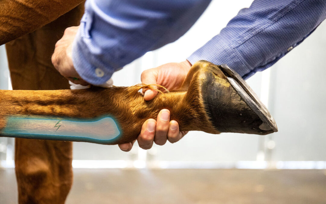
Lameness in horses is characterised by pain in the locomotor system. Visually, when moving, the transfer of body weight does not appear to be regular. In other words, its movements are asymmetrical. Even if lameness is common in horses, it is still difficult to diagnose, as its origin can be located either on one or more limbs, but also on the back or neck of the horse.
Detecting pathological asymmetry in its early form is one of the key issues in veterinary practice. This allows, besides other things, to optimise the treatment success through the implementation of optimal care.
But to what asymmetries can be linked? Are specific lamenesses more common in forelimbs? Or in hindlimbs? And how can they be identified?
Different kinds of lameness
Pathological equine locomotor asymmetries are lesions that can be classified into two types: osteoarticular and/or ligamentous and tendinous. There are several types of lameness in horses:
Abscess-related lameness
These are quite common and result from the penetration of a foreign body which leads to an abscess. The abscess is difficult to detect, which is why the diagnosis is often made when the locomotor asymmetry is visually noticeable. During the veterinary intervention, the abscess is pierced and a bandage is applied to help drain the pus and protect the wound. This operation does not usually leave any long-term sequelae.
Lameness related to laminitis
Laminitis is an inflammation of the lamina. In a horse, if most of the weight is on the forelimbs, horses with laminitis are more likely to shift their weight to the hindquarters to relieve pressure on the forelimbs, which are often the most affected. Lameness is often severe and the horse has great difficulty moving.
Lameness related to navicular syndrome
Lameness is usually chronic and often associated with pain that arises from the navicular bone and surrounding structures. Horses usually start limping between the ages of 7 and 9, but this can be variable. They frequently have short strides, and the asymmetry becomes more pronounced on hard ground.
Lameness associated with tendon or ligament injuries
They take their name from their origin: each of the horse’s limbs contains many major tendons and ligaments. The lameness is then caused by inflammation and/or partial or total rupture of the tendons. If the tendon is affected, it is called tendonitis. If the lameness is caused by inflammation of a ligament, it is referred to as desmitis. However, tendon or ligament lameness can also be the consequence of a badly treated wound: an infection in the sheath, septic tenosynovitis, or following a trauma.
Osteoarthritis-related lameness
Osteoarthritis is a degenerative joint disease characterised by the degradation of articulation. It is, in some cases, the result of natural age-related joint wear, but can also be associated with premature joint damage, caused for example by overtraining. Lameness is then caused by pain in the joint as well as structural changes due to cartilage destruction and bone remodelling.
Zoom on common lameness
Lameness is one of the most complex disorders to diagnose in the horse, as it can result from multiple anatomical sites and can be the consequence of many factors. However, some lameness is more common in the forelimbs – and some in the hindlimbs. This section is a simple overview of the most common lameness in the horse, according to the different limbs.
Forelimb lameness
Atrophy of the supraspinatus and infraspinatus muscles (sweeny) – Inflammation of the bursa of the biceps brachii (bicipital brucina) – Arthritis of the shoulder (scapulohumeral arthritis) – Paralysis of the radial nerve – Fracture of the ulna – Rupture of the medial collateral ligament of the humero-radial joint – Hygroma of the elbow Knee deviation towards the front (arch or brassicourt horse) – Knee deviation in the latero-medial plane in the foal – Hygroma of the knee – Traumatic arthritis of the knee – Intra-articular fracture of the carpal bones – Fracture of the accessory carpal bone – Rupture of the radial extensor of the carpus – Contracture of the finger flexor tendons – Tendonitis, Fractures of the proximal sepal bones – Sesamoiditis – Chip fractures of the proximal end of the first phalanx within the fetlock joint – Traumatic arthritis of the metacarpophalangeal joint Phalangeal forms – Osteitis of the extensor process (pyramidal eminence) of the third phalanx – Fracture of the extensor process (pyramidal eminence) of the third phalanx Cartilaginous javart – Cartilaginous forms – Laminitis – Navicular disease (podotrochleitis) – Fracture of the distal sesamoid bone – Fractures of the third phalanx – Osteitis of the foot – Penetrating wounds of the sole – Heel bruises and contusions of the sole
Hindlimbs lameness
Exercise-related myopathies – Myositis of the psoas and longissimus muscles – Overlapping thoracic and/or lumbar spinous processes – Sacroiliac subluxation – Pelvic fractures – Thrombosis of the caudal aorta or iliac arteries – Hip dislocation – Femoral head ligament rupture – Trochanteric bursitis – Femoral nerve palsy – Patella hooking – Chondromalacia of the patella – Stifle lameness – Ostéochondrose de la tubérosité tibiale – Fracture of the fibula – Rupture of the third peroneal muscle – Rupture of the hock cord – Rupture of the gastrocnemius muscle tendon – Fibrous myopathy and ossifying myopathy – Harper – Shivering – Bone sparing – Soft sparing – Venous sparing – Occult eparvin – Chip fractures of the tarsal tibial bone – Cession – Inflammation of the cuneus bursa branch – Guarding – Capelet – Flexor tendon laxity in the foal – Digital hyperextension in the foal – Toad
Forelimbs and hindlimbs lameness
Wobbler syndrome – Muscular dystrophy – Accidental section of the extensor tendons of the phalanges – Accidental section of the flexor tendons – Constriction of the annular ligament of the fetlock – Longitudinal fractures and comminuted fractures of the first phalanx – Fractures of the second phalanx – Forms due to rickets – Pincer, quarter and heel seines – Penetrating wounds of the white line – Fork rot – Wounds
Lameness Identification: the locomotor exam
To diagnose lameness, a veterinarian must have acute clinical and observational skills. It is essential to visually examine the horse, and the clinical examination is divided into two parts: a static examination followed by a dynamic examination.
The evaluation begins with a thorough physical examination and a detailed background check. The static examination assesses the horse’s conformation and posture visually. During the static examination, the veterinarian may use forceps to test the feet for tenderness that could be caused by navicular disease, as well as extension and flexion tests and palpation of the vertebrae for pain or heat.
Next, an assessment of the horse’s gait in movement is conducted. Lame horses have asymmetrical body movement due to an unconscious shift in body weight. Recognition of the resulting head nod and pelvic inclination is the basis for the diagnosis of lameness. The primary criteria for visual and objective assessment of lameness are asymmetries in the head movement for forelimb lameness and asymmetries in the pelvic movement for hindlimb lameness (Kramer et al. 2004).
During the dynamic examination, the lameness identification is enhanced by circular movements and flexions of the limbs. This examination is usually performed at trot: the horse’s locomotion is evaluated on different grounds, in a straight line and on a circle. It provides a first global view of the horse’s locomotion and can then indicate the sites that need to be investigated more precisely with radiographs, ultrasound, or other types of examinations. Finally, diagnostic anesthesia can also be performed to support the diagnosis of complex asymmetries, in order to confirm the exact location of the lesion.
Compensatory head nodding is most noticeable in horses with primary hindlimb lameness. This compensatory head movement asymmetry is easily misinterpreted as primary ipsilateral forelimb lameness (Kelmer and al, 2005).
According to Rhodin’s 2017 study, horses with induced hindlimb lameness and compensatory head movement asymmetry had head and withers movement asymmetries indicative of forelimbs lameness.
Wither movement symmetry can therefore be used to distinguish a head nod associated with true forelimb lameness from a compensatory head movement asymmetry caused by a forelimb asymmetry. Quantifying withers symmetry can therefore help to determine the location of the primary lameness, whether it is fore or hind limbs.

It is also possible, and more and more common, to perform a riding locomotor examination, at the 3 gaits. This approach allows a thorough evaluation of the horse’s locomotor system. By being monitored with veterinary technology, this examination allows a 360° analysis of the horse’s health.
Conclusion
Although lameness is a common pathology in the horse, its complexity makes it difficult to diagnose. Distinguishing between compensatory asymmetries and primary movement asymmetries directly related to pain is therefore a prerequisite for the successful evaluation of lameness.
Indeed, while lameness can be caused by two different types of lesions, it can be the consequence of many factors that, depending on their severity, will have more or less long-term consequences. For example, whether the horse is healthy or lame, the ground has a significant impact on its locomotion. As a result, it is critical to detect locomotor asymmetry as soon as possible. Locomotion quantification tools are fantastic diagnostic or longitudinal monitoring support for equine veterinarians!
SOURCES :
Rhodin, M., Persson-Sjodin, E., Egenvall, A., Serra Bragança, F.M., Pfau, T., Roepstorff, L., Weishaupt, M.A., Thomsen, M.H., van Weeren, P.R. and Hernlund, E. (2018). Vertical movement symmetry of the withers in horses with induced forelimb and hindlimb lameness at trot. Equine Veterinary Journal, 50(6), pp.818–824. doi:10.1111/evj.12844.
Weishaupt, M.A. (2008). Adaptation Strategies of Horses with Lameness. Veterinary Clinics of North America: Equine Practice, 24(1), pp.79–100. doi:10.1016/j.cveq.2007.11.010.
Keywords: equine locomotion, locomotor asymmetry, lameness, veterinary diagnosis, quantification tool, equine physiology, EnvA, CIRALE


