“In horses, there is a close relationship between cardiac capacity and athletic performance. Cardiac morphology and function evolve with training, which can lead to the appearance of arrhythmias or murmurs. A good knowledge of cardiac pathophysiology and a clear diagnostic approach are essential to determine the impact of abnormalities detected on cardiac auscultation on performance and to decide on the sports prognosis. Cardiac capacity may be impaired by abnormalities causing rhythm disorders or by valve insufficiencies that only manifest their effects during exercise. The practitioner can use useful complementary examinations including the stress test, the ECG and the echocardiography.”
How does the horse’s cardiovascular system work? What are the different existing cardiac pathologies? Do specific factors predispose to a certain cardiac pathology?
The horse’s cardiovascular system
The horse’s cardiovascular system is composed of the heart and blood vessels, which ensure blood circulation, transport of oxygen and elements of anti-infectious defences in the body but also allows the elimination of waste from cellular activity.

The horse’s heart is 25 to 30 cm long, has a diameter of about 20 cm and weighs on average 4 kg, which is about 1% of the horse’s mass. With each beat, the heart pumps blood and circulates it through the network of arteries and veins. Its resting heart rate – that is, the number of heartbeats per unit of time – averages 25 to 40 beats per minute and can reach 240 beats per minute during very intense effort.
The heart is divided into two independent parts called the right and left heart. The thoracic cage of the horse is flattened laterally which induces that the heart is flattened transversely and thus places the right heart in the cranial part and the left heart in the caudal part.
The heart has 4 cavities: the left atrium, the left ventricle, the right atrium and the right ventricle. The right side of the heart, consisting of the right atrium and ventricle, receives oxygen-depleted blood from the rest of the body. Conversely, the left side of the heart, consisting of the left atrium and left ventricle, receives freshly oxygenated blood from the lungs.

Cardiac pathologies in horses
Although the horse has a remarkable cardiovascular system, it can be subject to various cardiac pathologies:
- Amyloidosis: caused by the transformation of proteins that deposit and infiltrate the organs. When it affects the heart, it causes stiffness in the heart muscle and impairs its ability to relax and contract, leading to heart failure.
- Cardiac arrhythmias: abnormalities in heart rate, heart rhythm or conduction pattern due to abnormalities in impulse generation or conduction, or a combination of both.
Arrhythmias can be classified into two types:
- Bradyarrhythmias: can be physiological or pathological, sinus, nodal or due to atrioventricular block – Sinus bradycardia, Sinus arrhythmia, Sino-atrial block, Sino-atrial pause, Atrioventricular blocks.
- Tachyarrhythmias: occurs during atrial fibrillation, is characterised by irregular beating of the atria of the heart – Atrial Tachyarrhythmias, Ventricular Tachyarrhythmias.
- Cardiomyopathy: affects the heart muscle and reduces the heart’s ability to pump oxygen-rich blood to the rest of the body.
- Endocarditis: inflammation of the inner lining of the heart that may or may not be infectious. Infectious endocarditis is caused by bacteria and most often affects the heart valves.
- Aortic insufficiency: alteration of the structure of the valve which causes it to malfunction and, by not closing completely, allows blood to flow in the opposite direction. It is in fact a disturbance of the blood flow between the heart and the aorta.
- Mitral insufficiency: reflux of blood during the phase of contraction and ejection of the left ventricle towards the left atrium, is thus an anomaly of sealing between the atrium and the left ventricle.
- Congestive heart failure: inability of the heart to pump a sufficient amount of blood to meet the needs of the body. In older horses, it is mainly related to aortic insufficiency.
- Myocarditis: inflammation of the myocardium, which is the muscle tissue of the heart, develops various symptoms including tachycardia and tachypnea.
- Pericarditis: inflammation of the pericardium, the fibrous sac in which the heart and the roots of the large blood vessels are located.
- Heart murmur: an additional heart sound that can be heard with a stethoscope, which can be crescendo if it increases in intensity or decrescendo if it decreases in intensity.
What factors influence the risk of heart disease?
The horse’s life history can be key to diagnosis of a potential heart problem. Some conditions may affect a wider range of horses depending on age, breed, gender and the horse discipline.
AGE:
The probability of encountering congenital cardiovascular pathologies is more important in older horses. On the contrary, aortic regurgitation – leakage of the aortic valve: part of the blood ejected from the heart returns to the heart every time the left ventricle relaxes – is more often observed in young horses.
BREED:
Some heart conditions have a higher incidence in certain breeds, such as atrial fibrillation in heavy horses. Some congenital heart conditions are more common in the purebred Arabian, whereas there is a defect of the interventricular septum in miniature or pony horses. The Friesian, on the other hand, is more prone to aorto-pulmonary fistulas.
GENDER:
Stallions have a predisposition to develop fistulas – an abnormal channel between an artery and a vein – aortic, in which case the blood flows directly from an artery to a vein, without passing through the capillaries when it should flow from the arteries to the capillaries and then into the veins.
HORSE WORK:
During a sports horse examination, it is necessary to investigate whether the horse is suffering from exercise intolerance, but also to differentiate this from a possible genetic sports ability deficit or lack of training. If a effort test is carried out, the coach/trainer’s view of the way the horse is being worked is necessary in order to interpret the results of the test in the best possible way.

How to detect cardiac pathology?
An electrocardiogram or ECG is a veterinary measurement tool that records the electrical activity of the heart. It allows to control the cardiac function of a horse thanks to the retranscription of the electrical tracing of the heart. A veterinary examination with an ECG allows to analyse the heart rhythm and to detect possible cardiac pathologies.
For example, echocardiography allows the visualization of the cardiac anatomy and its possible abnormalities, while the electrocardiogram will give reliable and sure indications on the cardiac rhythm disorders. It can be used to investigate pathologies – in particular by analysing cardiac variability and the impact of stress on the cardiac rhythm – or to detect possible arrhythmias that may be linked to physical pain.
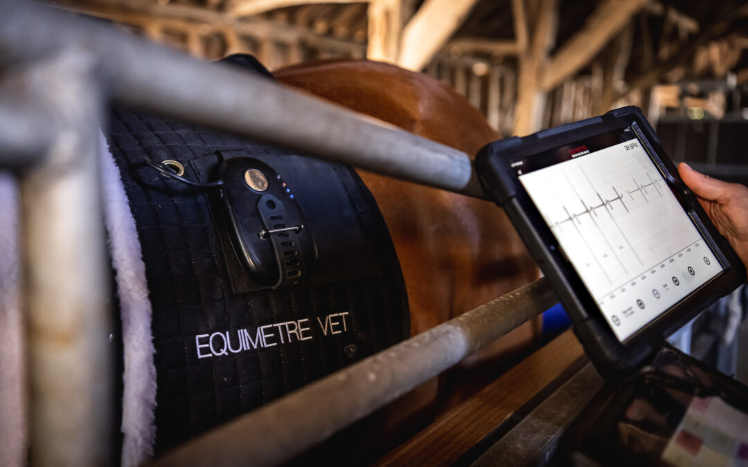
Collecting ECG during exercise with EQUIMETRE VET
EQUIMETRE VET collects the ECG at full speed with medical precision and simultaneously records the heart rate, locomotor and GPS data of each working horse. It has been scientifically validated twice, for heart rate and heart rate variability as well as for ECG quality and arrhythmia detection.
This access to a wealth of data allows research into all kinds of pathologies, both physiological and locomotor, to be oriented, investigated more quickly and monitored for rehabilitation. The use of data allows for the best possible protection of the health and safety of horses, even at a distance, thanks to the new opportunities offered by telemedicine.
CASE STUDY – Detect a mitral insufficiency with EQUIMETRE VET
Context: The case of Arion, a 2-year-old colt who had a promising first race but subsequently showed less physical progress than other 2-year-olds. We will now look at Arion’s data, collected during training. The red curve represents the heart rate, and the blue curve represents the speed.

What can we see? The HR stays at the maximum HR level even when the speed starts to decrease. This lag between the slowing down of the speed and the reduction of the HR is due to an oxygen deficit.
What can we see? Here, the longitudinal follow-up shows a degradation of the quality of recovery – studied thanks to the heart rate value after 15 minutes – which degrades over time.


ECG collected by EQUIMETRE VET during training.
After analysis of the EQUIMETRE VET data, an echocardiogram was performed and revealed mitral valve insufficiency. Arion will need to practice a less intense activity, or be retired.
Conclusion
The horse has a remarkable cardiovascular system, especially because of its great adaptability, but it is still subject to many cardiac pathologies. If some of these pathologies can be difficult to detect at their earliest stage, the veterinary diagnosis can be support by a tool that collects the ECG during exercise – such as EQUIMETRE VET – in order to analyse the variability of the horse’s heart rate over time.
SOURCES:
(2011) PLASTINATION D’UNE SERIE DE CŒURS DE CHEVAUX : OUTIL D’AIDE À LA COMPRÉHENSION DES IMAGES ECHOCARDIOGRAPHIQUES. Thesis. LA FACULTE DE MEDECINE DE CRETEIL.
Ouadah, C.Q. (no date) L’Appareil circulatoire… Le distributeur des métabolites et de l’oxygÚne. Available at: https://www.aucoeurdeschevaux.com/a/appareil-circulatoire-289/.
Delerue, M. (no date) Les principales maladies du cheval âgé. Available at: https://equipedia.ifce.fr/sante-et-bien-etre-animal/maladies/autres-maladies/les-principales-maladies-du-cheval-age/.
Comment fonctionne le cœur | Fondation des maladies du cœur et de l’AVC (no date). Available at: https://www.coeuretavc.ca/maladies-du-coeur/qu-est-ce-que-les-maladies-du-coeur/comment-fonctionne-le-coeur.
Le Point Vétérinaire.fr (no date) Cœur et performance : comment évaluer le système cardio-vasculaire chez le cheval athlète ? – Pratique Vétérinaire Equine n° 196 du 01/10/2017. Available at: https://www.lepointveterinaire.fr/publications/pratique-veterinaire-equine/article/n-196/coeur-et-performance-comment-evaluer-le-systeme-cardio-vasculaire-chez-le-cheval-athlete.html.
Weishaupt, M.A. (2008). Adaptation Strategies of Horses with Lameness. Veterinary Clinics of North America: Equine Practice, 24(1), pp.79–100. doi:10.1016/j.cveq.2007.11.010.
Keywords: equine cardiac pathologies, cardiovascular system, heart rate monitor, quantification tool, equine physiology, EnvA, CIRALE