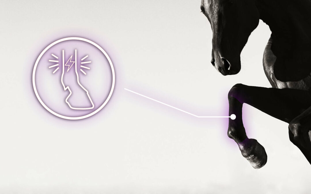
Lameness is a very common health problem for horses, and its diagnosis can be a real challenge for the veterinarian. The clinical evaluation of lameness is primarily based on the visual identification of asymmetrical movements, which are assumed to be pathological, as the horse displaces its body weight to discharge the painful structure(s).
Clinical evaluation: lameness diagnostic
Traditionally, veterinarians perform a methodical clinical evaluation on horses presenting with lameness. After taking a history of the lameness, the veterinarian performs a thorough examination of the musculoskeletal system of the static horse and then in motion. During the static examination, the veterinarian visually assesses the conformation and posture of the horse, palpates and mobilizes the relevant musculoskeletal structures. The horse’s locomotion is then assessed during a dynamic examination at a walk and trot in hand, in a straight line, but also during circle exercises on different surfaces and/or during ridden exercises. The veterinarian also performs flexion tests of the limbs to evaluate the potential aggravation of the pain, and therefore of the lameness, during this constrained mobilization. This way, he can determine the anatomical location of the painful structure.
To determine the anatomical source of the pain, he can finally proceed with diagnostic analgesia, using a local anesthetic to reduce or suppress the pain in a specific area of the limb, and then re-evaluating the horse to determine if the lameness has decreased.
The deductions made from all of this clinical examination (static and dynamic) then allow for an enlightened choice of different complementary examinations (X-ray, ultrasound, MRI, CT scan…), in an area of predilection, to make a lesion diagnosis. This means establishing the precise nature of the anatomical structure affected and the visible anomalies.
Identifying the lame limb(s)
Lameness evaluation is usually done at the walk and trot gaits. The trot, being a faster gait, involves more propulsion and lameness is usually more visible at this gait. This symmetrical gait is considered the easiest to detect asymmetrical movements.
Lame horses move asymmetrically because they are trying to shift some of their weight to relieve the painful structure(s). Asymmetry can refer to many aspects of movement, but in this context it is used to describe a difference in movement between the two “halves” of a trotting stride, or half stride. For example, an asymmetrical trot means that the horse moves differently when using one pair of diagonal limbs than when using the other.
What impact does lameness have on the horse’s locomotion?
For the symmetrical horse (considered healthy), the vertical displacement curves of the head, withers, and pelvis represent a perfect sinusoid. The two halves of the trotting stride are therefore similar: the movements of drop on one limb and then of vertical propulsion of the trunk are equivalent whether the horse uses its right or left limb.

Vertical displacement of the withers during a trot in one stride, posing Left Forelimb (LF) in pink and Right Forelimb (RF) in blue. Curves from the EQUISYM system.
Lameness leads horses to adapt their locomotor strategy in terms of vertical trunk movements, as well as a change in speed and limb synchronization. They reduce their stride length and speed.
Generally, lame horses adapt by taking shorter, faster strides: they increase the frequency of their strides and thus decrease their stride length. As a result, the limb propulsion per stride is reduced.
During the stride, the duration of the stance phase increases in horses with mild to moderate lameness, leaving the limb in contact with the ground for longer. This may appear counterintuitive, given the horse’s desire to avoid putting weight on the lame limb… On the other hand, the increased stance duration results in a reduction of the vertical amplitude by lowering the weight-bearing speed and propulsion.
Since the lame horse has a shorter stride duration but a longer stance duration, it follows that the swing or support phase of the stride is shortened. Changes in stride duration and swing phase are often more significant for horses with forelimb lameness than horses with hindlimb lameness.

CONCLUSION
Changes in the temporal parameters of the stride are usually helpful to understanding lameness. They are, however, highly dependent on the severity and type of lameness. As a result, the most widely used method for analyzing the movement changes caused by lameness is to observe changes in the vertical displacement of the horse’s head and trunk during a stride.
The development of quantification systems for locomotor asymmetries based on new sensor technologies allows to go further than visual observations by supplementing them with quantified and objective data on the horse’s locomotion. This is the case with the EQUISYM diagnostic tool, composed of 7 sensors installed on the horse (head, withers, pelvis, and 4 limbs) that communicate with a tablet to perform measurements and analyses in a simplified and autonomous way.
Discover how to quantify asymmetries in practice! EQUISYM assists veterinarians in evaluating locomotor asymmetries and diagnosing lameness.
Keywords: locomotor asymmetries, lameness, equine veterinarians, diagnosis, clinical examination
[1] Clayton HM. Chapter 14: Gait characteristics. In: The Dynamic Horse. Sport Horse Publications; 2004:178, 180.
[2] Deuel NR, Schamhardt HC, Merkens HW. Kinematics of induced reversible hind and fore hoof lamenesses in horses at the trot. Equine Vet J. 1995;(Suppl.18):147-151. doi:10.1111/j.2042-3306.1995.tb04908.x
[3] Weishaupt MA, Wiestner T, Hogg HP, Jordan P, Auer JA. Compensatory load redistribution of horses with induced weight-bearing forelimb lameness trotting on a treadmill. Vet J. 2006;171(1):135-146.
[4] Weishaupt MA, Wiestner T, Hogg HP, Jordan P, Auer JA. Compensatory load redistribution of horses with induced weightbearing hindlimb lameness trotting on a treadmill. Equine Vet J. 2004;36(8):727-733. doi:10.1016/j.tvjl.2004.09.004
[5] Buchner HHF, Savelberg HHCM, Schamhardt HC, Barneveld A. Temporal stride patterns in horses with experimentally induced fore‐ or hindlimb lameness. Equine Vet J. 1995;(Suppl. 18):161-165. doi:10.1111/j.2042- 3306.1995.tb04911.x
[6] Kallerud AS, Someting in the way you move: Studies of movement asymmetry in young Standardbred trotters. PhD Thesis. ISSN 1894-6402. 2021.