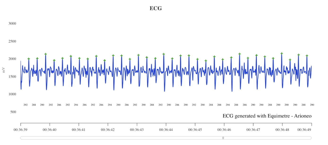
The electrocardiogram (ECG) is an essential tool for detecting cardiac pathologies in horses. It is a reliable, non-invasive, and easy-to-use technique that allows for the investigation of cardiac rhythm abnormalities.
With the Dashboard Vet platform, veterinarians can analyze in detail the different arrhythmias and electrical anomalies of the heart. This guide aims to present the main cardiac pathologies detectable on an ECG, with visual examples taken from the platform.
On the agenda in this article:
- Normal ECG: Understanding the key components
- Detection of cardiac arrhythmias
- Distinguishing noise from an arrhythmia
Discover the presentation video of our Dashboard Vet platform: watch it here
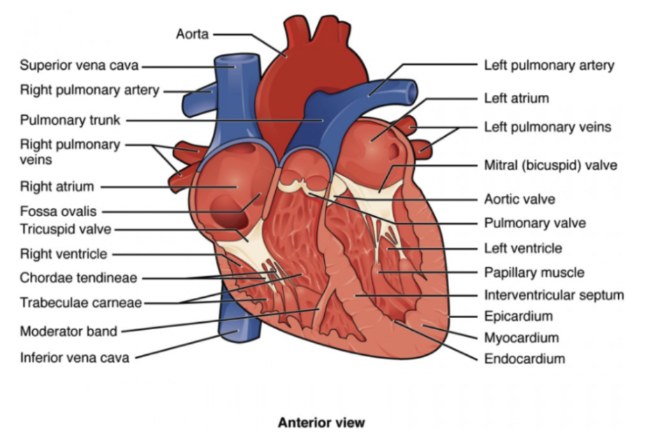
Diagram of the horse’s heart
Normal ECG: Understanding the key components
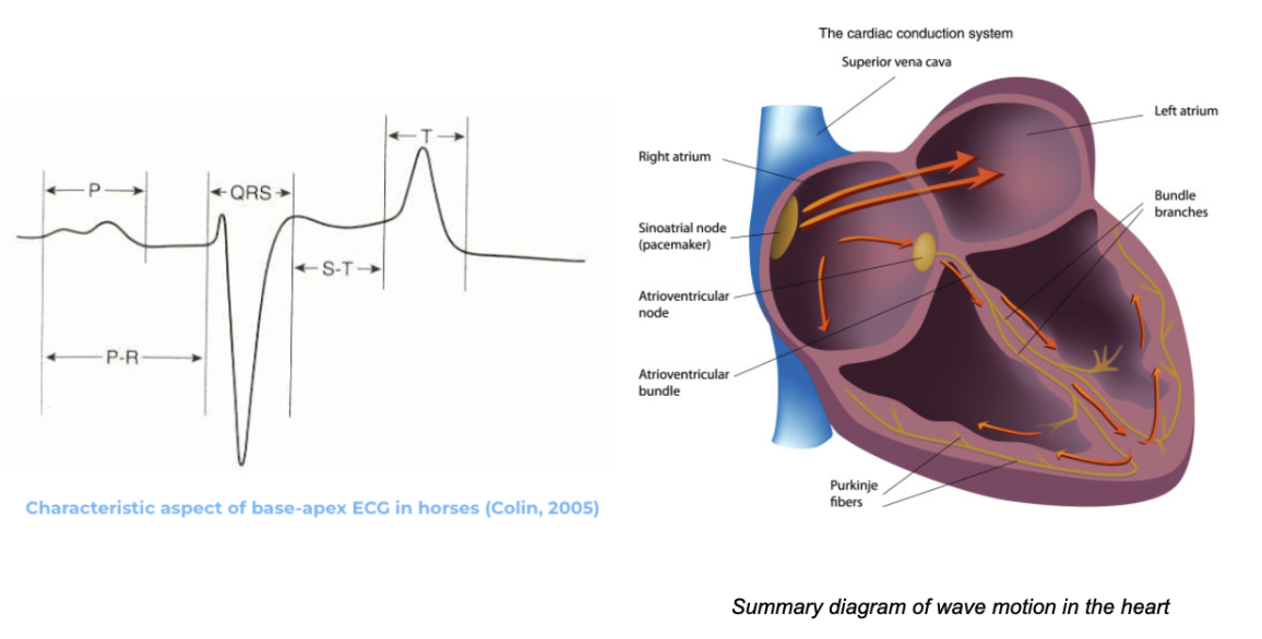
Before analyzing arrhythmias, it is essential to understand what constitutes a normal ECG:
- P wave: Represents atrial depolarization, meaning the electrical activation that initiates the heart’s contraction.
✅ Start of the cardiac cycle: It precedes the QRS complex.
✅ Short duration: Approximately 0.08 to 0.10 seconds in horses.
✅ Low stride length: Often less than 0.2 mV.
✅ Normal morphology: Rounded, irregular, or bifid, without interruption.
- QRS Complex: Represents the electrical activity associated with ventricular depolarization. It is one of the key elements in ECG interpretation, allowing for the analysis of electrical conduction within the ventricles. The QRS complex generally consists of three waves, whose morphology may vary depending on the horse and electrode placement:
- Q wave: The first negative wave, which is often absent in horses. Typically, an RS complex is seen, but for convenience and in line with human medical terminology, it is commonly referred to as the QRS complex.
- R wave: The first positive wave.
- S wave: The second negative wave.

The duration of the QRS complex reflects the time required for depolarization to propagate through the ventricular myocardium. It ranges from 0.08 to 0.16 seconds (Sevestre, 1972).
- T wave: Represents the repolarization of the cardiac ventricles. It immediately follows the QRS complex and is a key component in recording the electrical activity of the horse’s heart.
Appearance of the T wave: It is generally positive in horses in standard derivations (i.e., it is directed upwards on the ECG). However, in some derivations, it may be negative or biphasic (both positive and negative).
Duration and shape: The T wave is usually broader than the P wave and can vary in stride length. It is generally higher and more rounded in a healthy horse, but its appearance may be influenced by hydration status, stress, or other factors.
- RR interval: Represents the distance between two successive R wave peaks, that is, between two QRS complexes. This interval corresponds to the time between two consecutive heartbeats.
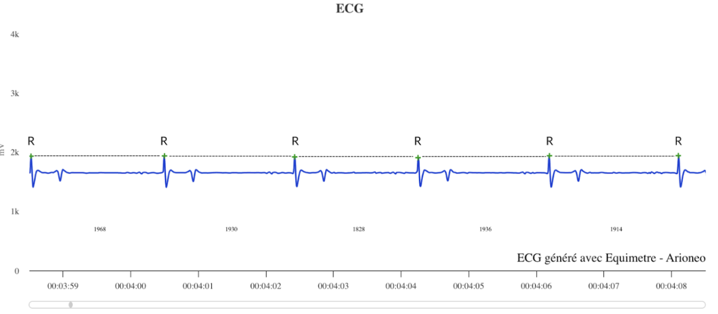
The RR interval in horses allows for the measurement and calculation of heart rate. A short interval indicates a faster rhythm, while a long interval indicates a slower rhythm. Heart rate can be calculated by dividing 60 by the duration of the RR interval in seconds. For example, an RR interval of 0.6 seconds corresponds to a heart rate of 100 bpm (60 / 0.6 = 100 bpm).
At rest, the average heart rate of an adult horse ranges from 32 to 44 beats per minute, while for a foal in his first weeks of life, it ranges from 50 to 70 beats per minute. However, this can vary depending on the individual horse (size, breed, temperament) and external factors (exercise, stress).
Detection of cardiac arrhythmias
The main indicator of an arrhythmia is an irregularity in the frequency or morphology of the QRS complex. Abnormal variations in the duration of RR intervals, whether short or long, can indicate cardiac rhythm disorders that require thorough clinical evaluation. Analyzing these irregularities is essential, as some arrhythmias—potentially serious—can occur both at rest and during exercise. Early detection enables appropriate management and reduces the risk of cardiovascular complications. The Dashboard Vet platform, in combination with EQUIMETRE recordings, allows for the automatic detection of RR intervals, making diagnosis easier.
Normal ECG at over 200 bpm collected with EQUIMETRE.
Here are the main arrhythmias that can be identified using ECG.
1. Physiological arrhythmias detectable at rest
a) Bradycardias – Atrioventricular (AV) Blocks
A bradycardia is characterized by an abnormally slow heart rate. There are different types of bradycardias:
- First-degree AV block: This is a slowing of conduction between the atria and the ventricles (there is no missed beat, only a prolongation of the PR interval—the delay between the P wave and the QRS complex). There are no clinical signs; it is generally asymptomatic and benign, with no known impact on performance.
Possible causes:
A first-degree AV block can be physiological in origin, typically due to marked vagal tone (increased parasympathetic activity), which is commonly seen in athletic horses or those at rest. This phenomenon is very frequent in horses in good physical condition.
This is similar to a “sinusal pause,” except that during a sinusal pause, there is no P wave—meaning no atrial depolarization.
2. Pathological arrhythmias detectable at rest
a) Ventricular tachycardia
Ventricular tachycardia (VT) is an arrhythmia characterized by a rapid succession of abnormal ventricular beats, compromising cardiac efficiency. The electrical impulses originate from the ventricles rather than the normal sinoatrial node.
On the electrocardiogram (ECG), ventricular tachycardia is manifested by:
- A rapid resting heart rate (often >100–120 bpm in horses).
- Wide and abnormal QRS complexes on the ECG (since activation does not follow the normal conduction pathway).
- Possible atrioventricular dissociation (the atria and ventricles beat independently).
Causes:
This arrhythmia can be caused by a variety of factors, both cardiac and non-cardiac. Cardiac causes include myocarditis or myocardial fibrosis, dysplasia, or congenital abnormalities of the cardiac tissue. Non-cardiac causes may be related to electrolyte imbalances, thoracic trauma, toxins, severe hypoxia, or sepsis.
Treatment:
When ventricular tachycardia is detected at rest, it is crucial to assess his heart rate, duration, and clinical effects (peripheral perfusion, blood pressure, signs of weakness). Even at rest, ventricular tachycardia is never considered normal and indicates potentially serious electrical instability.
The primary treatment is the administration of an antiarrhythmic. Lidocaine is the first-line medication: it is administered intravenously as a bolus (usually 1.3 mg/kg). If the response is positive (slowing or disappearance of tachycardia), a continuous lidocaine infusion can be started to stabilize the horse.
b) Bradycardias – Atrioventricular (AV) Blocks
In the case of a pathological origin, it may be due to electrolyte disturbances (hypokalemia—low potassium levels in the blood—or hypocalcemia, for example—too low calcium levels), side effects of medication, inflammation, or degeneration of nodal tissue (myocarditis, cardiac fibrosis).
Treatment:
Most of the time, first-degree AV block is physiological and does not require any treatment, as the block generally disappears as soon as the horse starts moving. In the case of a pathological origin, the underlying cause mentioned above should be treated and regular ECG monitoring should be adopted to monitor progression.
- Second-degree AV block: Some electrical impulses are blocked before reaching the ventricles, resulting in intermittent absence of ventricular contraction. Presence of P waves not followed by a QRS complex.
Additional information: In first-degree AV block, the horse only misses one beat. In second-degree AV block, generally one beat is missed between two normal beats (there is a P wave, then a pause, and no QRS for two beats).
- Physiological if it disappears with effort or stimulation
- Pathological if it persists
Possible causes:
There are several possible causes similar to those of first-degree AV block, such as electrolyte disturbances, drug intoxication, or underlying heart disease. It can also be due to systemic pathologies (sepsis, digestive disorders).
Treatments:
Treatments therefore depend on the different causes, but it is possible to correct the various metabolic imbalances with medical treatment or by placing a pacemaker.
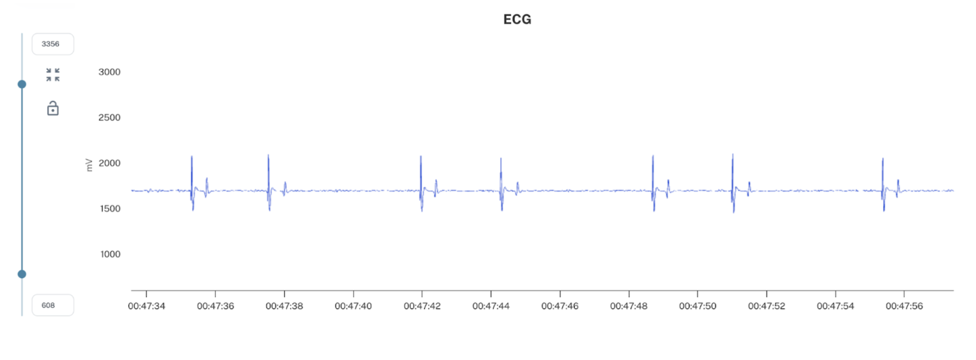
This ECG shows second-degree AV blocks recorded with EQUIMETRE
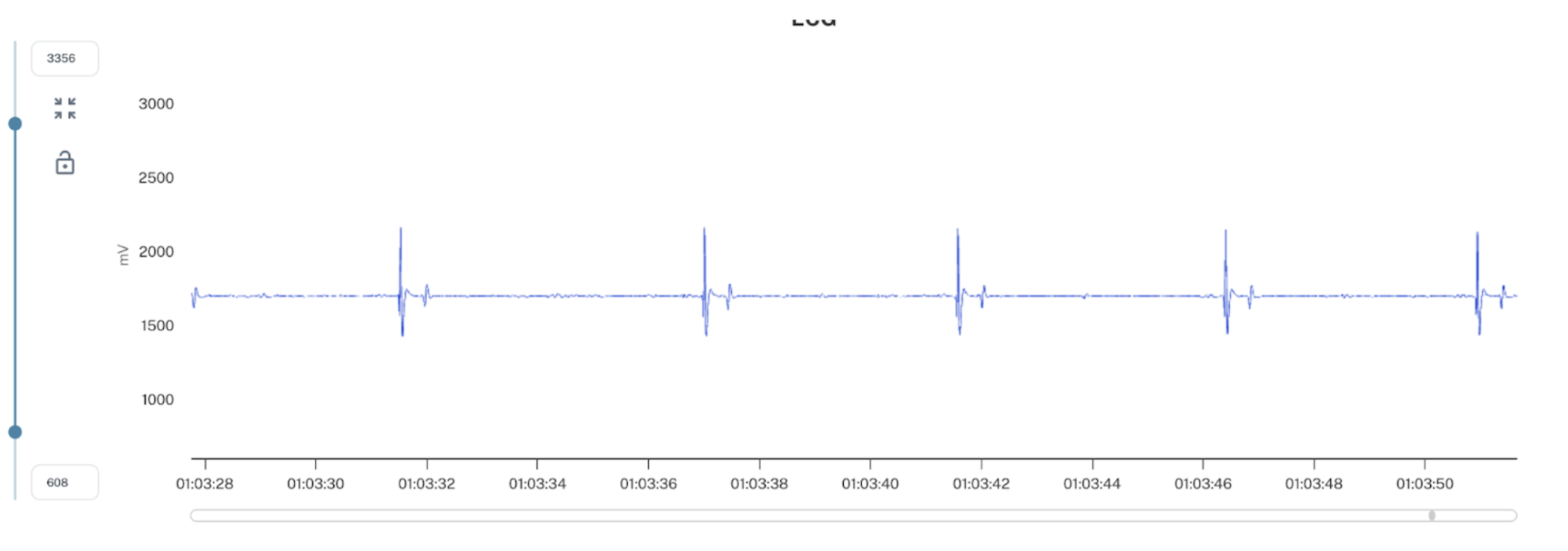
This ECG shows a block at every beat recorded with EQUIMETRE
- Third-degree AV block: The electrical signal no longer passes from the atria to the ventricles, so the ventricles beat on their own, not following the atria. P waves are not related to QRS complexes. Slow and regular heart rate. Severe bradycardia (< 20 bpm). In this case, there is incompatibility with performance and a risk of syncope.
Causes :
Third-degree AV blocks induce more severe cardiac pathologies such as idiopathic fibrosis of the conduction system, ischemic or inflammatory heart diseases (myocarditis, endocarditis).
Treatments:
To treat these pathologies, it is possible to place a pacemaker in case of symptoms (syncope) or critical ventricular rate. In milder cases, a problem-oriented treatment is appropriate, for example anti-inflammatories for myocarditis.
AV blocks are common arrhythmias in horses, some of which are benign and physiological, while others require serious management. An exercising ECG can also be used to differentiate a benign block from a pathological block. The most severe forms can compromise the horse’s ability to perform at a high level.
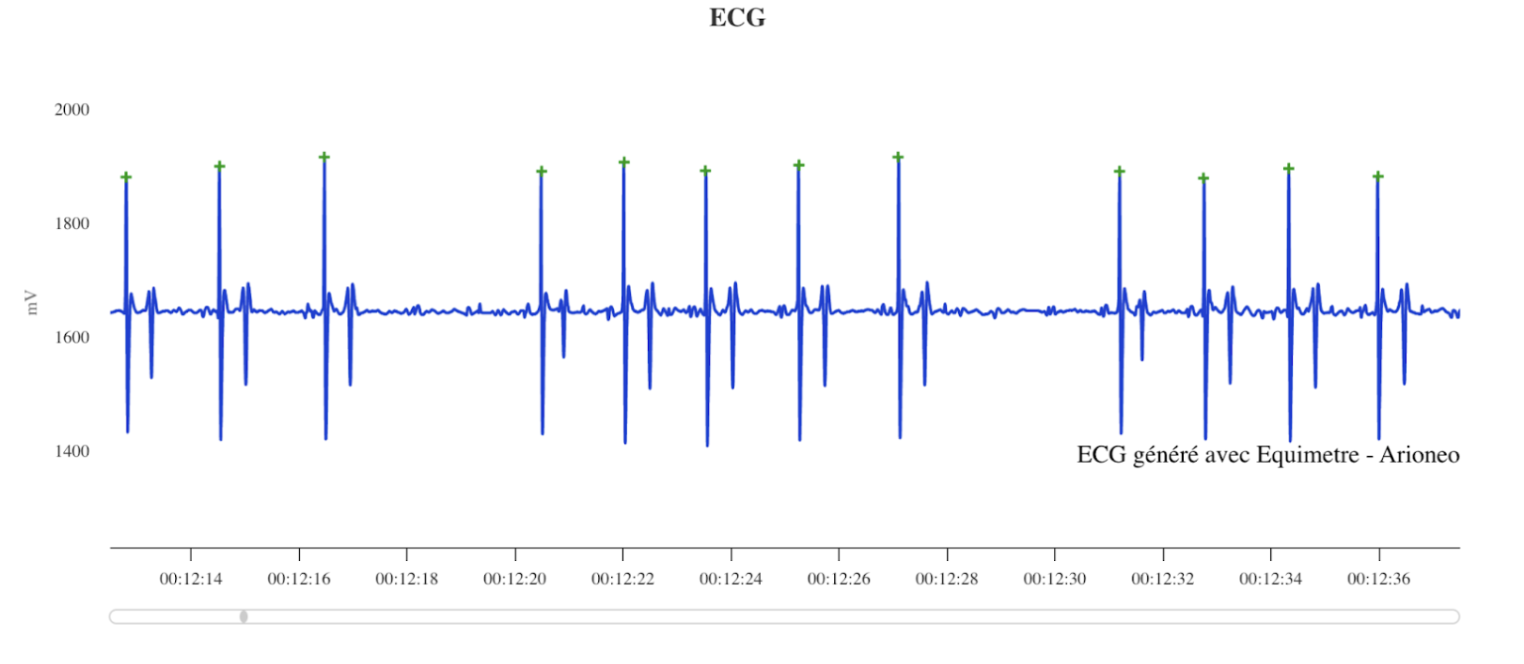
This ECG shows sinusal pauses recorded with EQUIMETRE
3) Pathological arrhythmias detectable during exercise
a) Atrial fibrillation
Atrial fibrillation is characterized by an irregularly irregular rhythm that can impair performance, increase the risk of sudden death, and may contribute to EIPH (Exercise-Induced Pulmonary Hemorrhage). This arrhythmia affects the performance of horses. It presents as rapid beats interspersed with pauses of variable duration when the heart rate remains normal. This arrhythmia can occur with or without underlying cardiac pathology, thus requiring echocardiography to identify the cause. It is common in sport horses, especially those of larger size.
It is not recommended for a horse with atrial fibrillation to undertake intense exercise (racing, galloping, etc.) due to the high risk of collapse (a sudden fall or episode of weakness, often caused by a temporary loss of effective cardiac output). For sport horses engaged in less intense or leisure activities, regular monitoring with exercise ECG is strongly recommended.
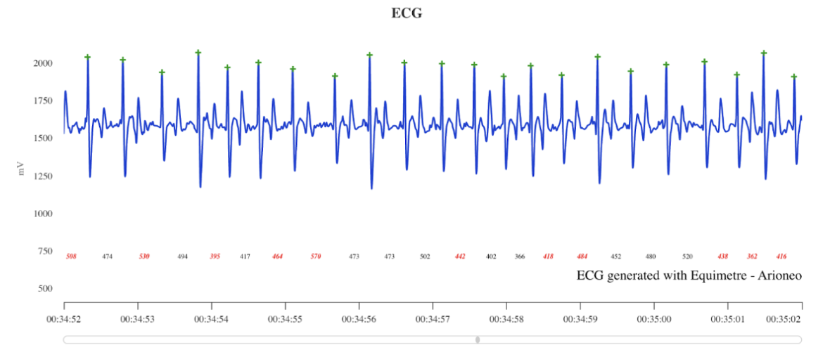
This ECG shows atrial fibrillation recorded with EQUIMETRE
Clinical significance:
📌 An abnormal P wave may indicate atrial hypertrophy or a disturbance in electrical conduction between the atria and the ventricles.
📌 In cases of atrial fibrillation, the P wave may be absent and replaced by irregular oscillations.
Diagnostic precaution: Before initiating treatment, it is essential to rule out an underlying pathology such as mitral or tricuspid valve regurgitation. These valvular diseases can be detected clinically by the presence of a murmur on auscultation.
Causes:
Atrial fibrillation can be caused by abnormal increases in heart size, known as cardiomegaly, or structural changes in the atria, electrolyte imbalances, intense exercise, or stress.
Treatments:
Transvenous electrical cardioversion (TVEC) is currently the preferred treatment. TVEC involves placing electrodes, via catheters inserted through the jugular vein, near the atrial tissue in the heart. A biphasic electrical shock is then delivered under general anesthesia, causing complete depolarization of the atrial myocardium. This instantly restores a normal sinus rhythm by interrupting the fibrillation. This treatment is not exceptional and can be performed routinely. This method is preferred over medical treatments, which can also be used but have a high potential toxicity.
b) Atrial tachycardia
Atrial tachycardia is a rare arrhythmia in horses, characterized by a rapid and regular atrial rate without the electrical disorganization seen in atrial fibrillation.
Cause:
It may be transient or persistent, secondary to heart disease, electrolyte imbalance, systemic disorder, stress, anesthesia, or idiopathic. Clinical signs include exercise intolerance, elevated resting heart rate, sometimes without other symptoms. Diagnosis is based on ECG, supplemented by echocardiography and blood tests.
Treatment:
Treatment targets the underlying cause; antiarrhythmic drugs are rarely used, and electrical cardioversion is reserved for exceptional cases. The prognosis depends on the cause and the chronicity of the arrhythmia.
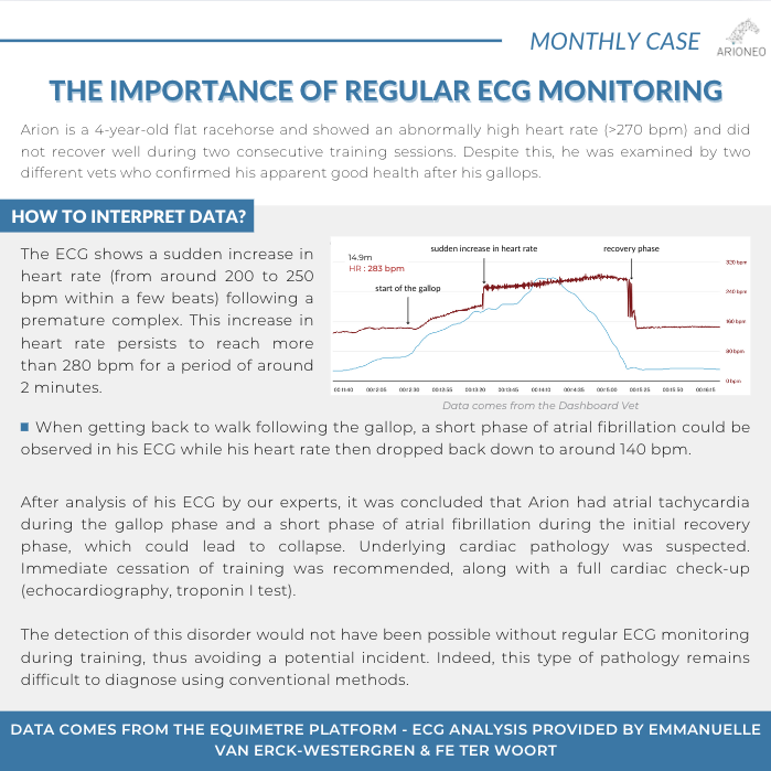
c) Premature complexes
Premature complexes are abnormal heartbeats that occur earlier than expected in the normal cardiac cycle. They are caused by premature electrical activation originating from an abnormal site in the heart, either in the atria (supraventricular premature complexes) or in the ventricles (ventricular premature complexes).
These arrhythmias can be transient or pathological, potentially affecting the horse’s performance and, in some cases, posing a safety risk. According to an ongoing study in Hong Kong, any arrhythmia detected during exercise (even isolated) is associated with an increased risk of poor performance. It may be a sign of an underlying pathology, even if it appears isolated. It is not normal to detect premature complexes at any time during a recording, including the initial recovery phase.
Distinction to make: A sinusal arrhythmia can be physiological: it reflects variations in sympathetic/parasympathetic tone and is therefore frequent and normal during recovery. In contrast, premature complexes (supraventricular or ventricular) are not, even during recovery.
- Supraventricular Premature Complexes (SPCs): A premature QRS complex originating from an ectopic focus in the atria. It is characterized by a premature P wave, a QRS complex of normal morphology, and an incomplete compensatory pause.
- Ventricular Premature Complexes (VPCs): A premature QRS complex originating from an ectopic focus in the ventricles. It is characterized by the absence of an associated P wave (since the impulse does not originate from the sinus node) and a complete compensatory pause.
It is important to note that the presence of a premature complex is never entirely normal, but it is not uncommon to observe them occasionally, especially at the beginning of the recovery phase. If the number of premature complexes remains less than or equal to 5 during intense exercise, this is generally not concerning but should still be monitored. However, more than 5 premature complexes is a sign of abnormality requiring thorough investigation and increased monitoring. The higher the number of premature complexes, the greater the risk of an underlying rhythm disorder.
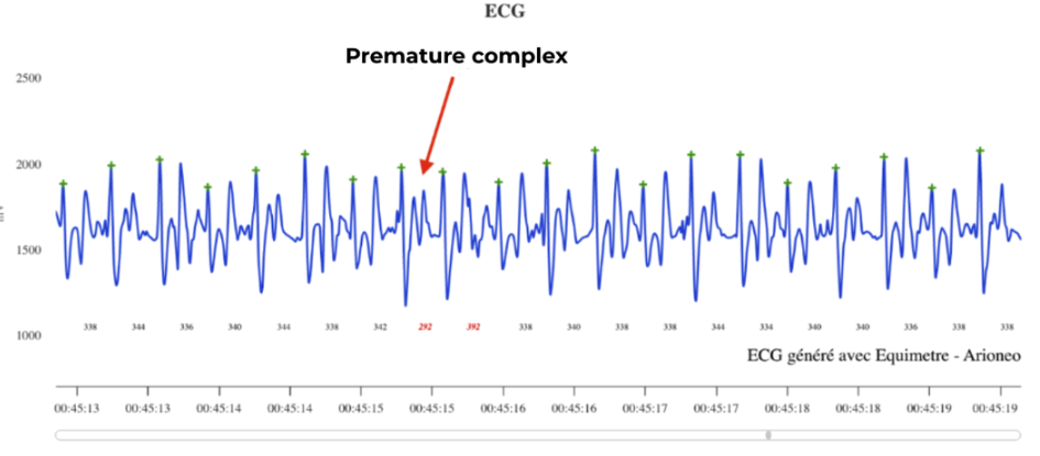
This ECG shows premature complexes recorded with EQUIMETRE
d) Complex arrhythmias: bigeminy, couplets, triplets, and others
Bigeminy is a specific form of arrhythmia in which heartbeats appear in pairs, alternating a normal beat (sinus complex) and a premature beat (extrasystole). This type of arrhythmia can be of ventricular or supraventricular origin. Couplets and triplets represent another form of complex arrhythmia, characterized by the consecutive occurrence of two (couplet) or three (triplet) premature beats. Their presence reflects greater electrical instability of the myocardium and, like bigeminy, may be of supraventricular or ventricular origin. The longer the sequence of premature beats, the higher the risk of progression to severe rhythm disturbances.
These different arrhythmias share common causes, such as electrolyte imbalances (hypokalemia, hypomagnesemia), heart diseases (myocarditis, fibrosis, valvular disease), respiratory pathologies, stress, intense exercise, or the action of toxins or certain medications.
Treatment primarily targets correction of the underlying cause: rebalancing electrolytes, treating an infection, discontinuing a causative medication, and strict rest, especially if the arrhythmia occurs during exercise.
In more severe cases, especially in the presence of frequent ventricular premature complexes or clinical signs, antiarrhythmic drugs may be administered under veterinary supervision, with intravenous lidocaine being the reference treatment for controlling complex arrhythmias. In all cases, regular ECG monitoring is essential to assess progression and adapt management.
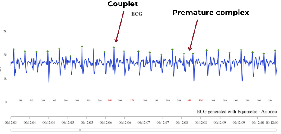
example of a couplet followed by a premature complex recorded with EQUIMETRE
Distinguishing noise from an arrhythmia
It is essential to differentiate a true cardiac abnormality from “noise” caused by poor electrode placement or horse movement. Analyzing an electrocardiogram (ECG) in horses can sometimes be complicated due to noise, which may falsely resemble an arrhythmia. Here are the key elements to distinguish them:
✔ Example of noise:
- Irregular tracing with extraneous spikes lacking logical structure.
- The problem disappears when the electrode position is changed.
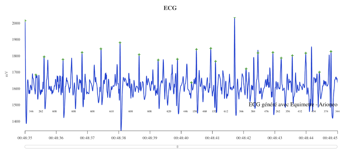
Impossible to distinguish the P waves, QRS complexes, and T waves.
By integrating exercise ECG into veterinary monitoring, it becomes possible to detect cardiac abnormalities early and optimize the management of athletic horses. Thanks to EQUIMETRE, veterinarians can remotely monitor the cardiac evolution of horses, thus ensuring regular follow-up, especially for those suffering from cardiac pathologies. Its simple and intuitive use allows it to be easily integrated into a care routine, offering reliable monitoring without constraints. In addition, with the Dashboard Vet, all physiological and ECG data are centralized and analyzed on a single platform, making interpretation, sharing with the veterinarian, and decision-making easier. A real asset for preventive and precision sports medicine!
Photo credit: Charlotte Majed.
Sources :
- https://www2.vetagro-sup.fr/bib/fondoc/th_sout/th_pdf/2005lyon124.pdf
- https://mediatheque.ifce.fr/doc_num.php?explnum_id=22451
- https://www.cliniqueequinedemeslay.com/departments/cardiologie/#:~:text=Lorsqu’une%20arythmie%20cardiaque%2C%20une,de%20diagnostiquer%20une%20%C3%A9ventuelle%20anomalie.
- https://fac.umc.edu.dz/vet/Cours_Ligne/cours_21_22/Patho_Equine/proedeutique_sys_cardio_resp.pdf
- https://orbi.uliege.be/bitstream/2268/123716/1/AMORY_ARYTHMIES.pdf
- https://www.vetopedia.fr/arythmies-cardiaques/
- https://baillyveterinaires.com/lelectrocardiogramme-chez-le-cheval/
- https://www.nagwa.com/fr/explainers/912123271719/
- https://orbi.uliege.be/bitstream/2268/123716/1/AMORY_ARYTHMIES.pdf
- https://www.lepointveterinaire.fr/publications/pratique-veterinaire-equine/article/n-154/traitement-transitoire-d-une-tachycardie-ventriculaire-paroxystique-par-la-phenytoine.html
- https://www.msdmanuals.com/fr/professional/troubles-cardiovasculaires/arythmies-cardiaques-sp%C3%A9cifiques/bloc-auriculoventriculaire


